Anatomical considerations and continence following prostate surgery

Prostate surgery may be undertaken to treat prostate cancer or benign prostatic hyperplasia (BPH), using open, laparoscopic, robotic or endoscopic techniques. The anatomical structures affected, and the resulting risk of urinary incontinence, vary according to the procedure performed. This article focuses principally on continence following radical prostatectomy for prostate cancer, where preservation of the urethral sphincter complex, its supporting structures and associated neural pathways may influence postoperative continence recovery.

Anatomical considerations to reduce incontinence


Continence depends on the coordinated function of several interconnected anatomical components, as follows:

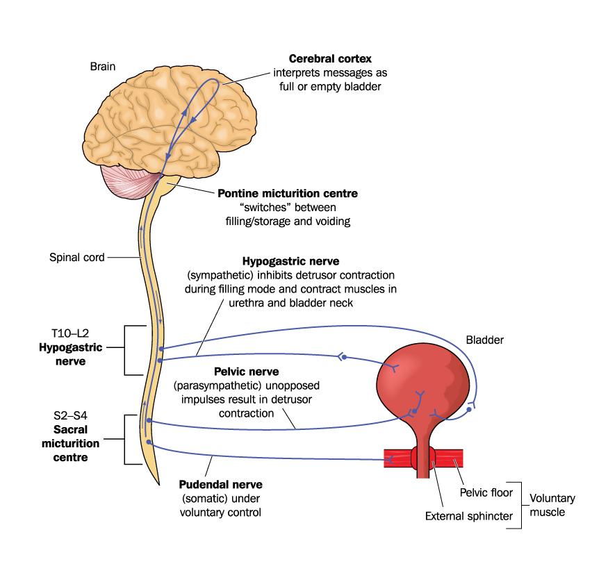
Neural pathways
  • Pudendal nerve and its branches
  • Pelvic autonomic nerves and the inferior hypogastric plexus.
Urethral sphincter complex and pelvic floor musculature
  • Internal and external urethral sphincter components
  • Membranous urethra and its surrounding musculature
  • Levator ani and puboperinealis muscles.
Fascial and ligamentous support
  • Detrusor apron and anterior attachments
  • Endopelvic and periprostatic fasciae
  • Denonvilliers’ fascia and associated posterior support
  • Arcus tendineus and related lateral support.
Bony support
  • Pubic bone and the structures attaching the continence mechanism to it.
A recent updated anatomical review describes the external and internal sphincters together with the membranous urethra as components of the urethral sphincter complex, supporting this more functional classification (Mandel et al, 2026).

Figure 1. Nervous control of the urinary bladder via the hypogastric, pelvic and pudendal nerves. Credit: Blamb/Shutterstock

Nerve-sparing surgery


Following improved understanding of the neurovascular anatomy, nerve-sparing approaches were introduced to preserve neural structures where this is oncologically appropriate. NeuroSAFE uses intraoperative frozen-section examination of tissue adjacent to the neurovascular structures to support decisions about nerve preservation. In a large observational study conducted after the introduction of NeuroSAFE, 92% of respondents at one year and 94% at two years reported using no more than one pad per day. These findings are encouraging, although they should not be interpreted as proving that NeuroSAFE alone caused the reported continence outcomes (van der Slot et al, 2023).

Preserving fibrous structures


In a prospective single-centre study of 311 patients undergoing Retzius-sparing robot-assisted radical prostatectomy, greater preservation of the detrusor apron was associated with improved continence recovery. Sacrifice of the detrusor apron was associated with delayed recovery and a higher incidence of incontinence. These findings support preservation where surgically and oncologically appropriate, although their applicability to other prostatectomy approaches requires further evaluation (Kishore et al, 2025).

Muscles affected by prostate surgery


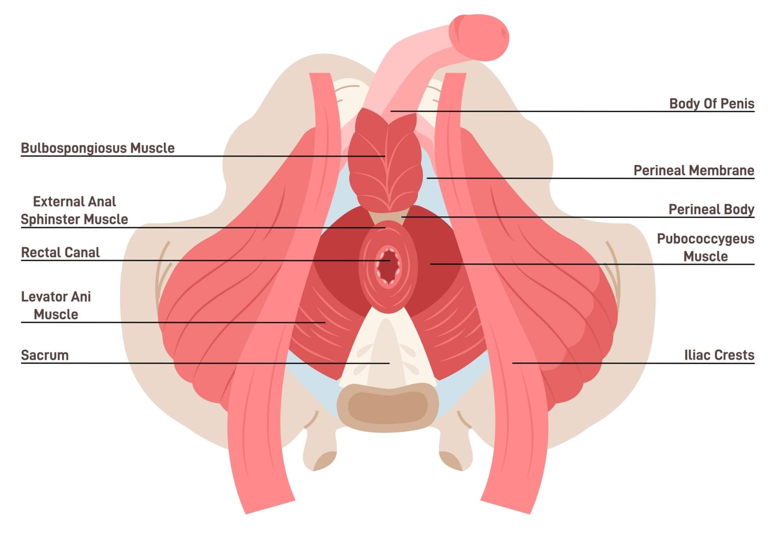
Prostate surgery may affect the pelvic floor musculature and the urethral sphincter complex. The pelvic floor supports the bladder and urethra, while coordinated activity of the smooth and striated components of the urethral sphincter mechanism contributes to urine storage and voluntary continence.


The importance of the urethra


Post-prostatectomy incontinence is commonly associated with impairment of the urethral sphincter mechanism. Contributing factors may include reduced functional urethral length, neural injury, direct muscle damage and loss of surrounding support. Greater membranous urethral length has been associated with improved continence recovery after radical prostatectomy. Careful apical dissection aims to preserve as much functional urethral length as is surgically and oncologically appropriate (Barakat et al, 2024)

Figure 2. Pelvic floor muscles in men. Credit: inspiring.team/Shutterstock

Conclusions


Continence following radical prostatectomy depends on the coordinated function of the urethral sphincter complex, pelvic floor, neural pathways and surrounding fascial and ligamentous support. Evidence suggests that preservation of membranous urethral length, neural structures, the detrusor apron and selected fascial supports may contribute to earlier or improved continence recovery. However, the techniques used must be tailored to the individual patient and balanced against the overriding need for oncological safety.

References


Barakat B, Addali M, Hadaschik B, Rehme C, Hijazi S, Zaqout S (2024) Predictors of early continence recovery following radical prostatectomy, including transperineal ultrasound to evaluate the membranous urethra length (CHECK-MUL Study). Diagnostics (Basel) 14(8): 853

Kishore TA, Mathew J, Boppanna VB, Fellow S, Prabhakaran S (2025) Impact of graded detrusor preservation on continence in Retzius-sparing robotic-assisted prostatectomy. World J Urol 43(1): 150

Mandel A, Parekh S, Choudhary M, Bessede T, Walz J, Tewari A (2026) Analysis of the current surgical anatomical knowledge of radical prostatectomy: an updated review. Eur Urol 89(2): 128–39. Available online: www.europeanurology.com/article/S0302-2838(25)00344-6/abstract

van der Slot MA, Remmers S, van Leenders GJLH et al; Anser Prostate Cancer Network (2023) Urinary incontinence and sexual function after the introduction of NeuroSAFE in radical prostatectomy for prostate cancer. Eur Urol Focus 9(5): 824–31