What can you learn from lab research findings and how might they relate to your clinical practice?

Introduction


This article looks at how you can assess different types of research papers and apply appropriate parts of their results to your practice and patients.
 
As we’ve covered in previous articles on the CLP, there are many different types of research, from the in vitro, in vivo and ex vivo clinical studies undertaken in laboratories, to qualitative studies of patients’ perspectives on a treatment, condition or situation, to large-scale randomised, controlled trials of new treatments or medicines, that are considered to be the ‘gold standard’ of the evidence hierarchy. All these approaches have their place in healthcare research. In the current era of evidence-based practice, however, it is important for clinical decision-makers to appreciate the strengths and limitations of different types of research evidence, and how to apply findings as part of evidence-based decision-making in clinical practice. Here, we present a case study of some recent research and highlight its relevance.

Research paper


For the purposes of this article, we are going to look at a new piece of research undertaken by researchers from the School of Pharmacy, Queen’s University Belfast, Belfast and Convatec, and funded by Convatec.

This looked at the development and optimisation of an ex vivo porcine urethral model for investigating intermittent catheter-associated urethral microtrauma. Research has shown that male porcine urethras are excellent tissue models for male human urethras (Tissue Source, 2020; André et al, 2022; Kazi et al, 2025).

The research involved taking urethras out of male pigs that had been medically euthanised, and then using a range of techniques in the laboratory to look at the effects that a range of different catheters had on various aspects. These included: the effect on the lining of the urethra, measuring the friction and force required to insert and remove the catheter, looking at how much the catheters dried out the lining of the urethra, and also how much of the catheter coating was left behind in the urethra once the catheter had been removed. You can read the full paper (Burns et al, 2025) at https://doi.org/10.1016/j.matdes.2025.114727

 
Urinary Retention Fowlers Syndrome

Key findings

Force needed to remove catheters

The mean total work done (force used to remove the catheter) increased on withdrawal for all the hydrophilic polyvinylpyrrolidone (PVP)-coated catheters compared to that needed for insertion, whereas the mean total work done on withdrawal of the integrated amphiphilic surfactant (IAS) catheter was significantly decreased compared to that used for insertion. This difference could suggest that the hydrophilic PVP-coated catheters undergo partial dry-out leading to mucoadhesion between the catheter surface and urethral tissue, requiring greater force to remove them from the model. Previous work by this team (Pollard et al, 2022) found that the force needed to remove PVP-coated catheters was significantly more than the force needed to remove an IAS catheter in an in vitro model. It is unclear how these forces measured ex vivo compare to the forces needed to remove catheters in humans, but this is currently the best evidence available because of the ethics of doing the study in a human population.
 
Effect on uroepithelial membrane

To assess the effect of intermittent catheterisation on urethral microtrauma, 4 µm sections of urethra were studied post-catheterisation for evidence of microtrauma. Damage to the urethral transitional membrane was assessed by staining and then examination under a microscope. All intermittent catheters exerted some degree of damage on the transitional membrane. Both the IAS catheter and the PVP control showed comparable results to the negative control, which was an uncatheterised urethra, meaning that the PVP solution and the IAS did not affect the staining or microscope results on their own. All hydrophilic PVP-coated catheters displayed significantly less fluorescence intensity than the negative control, indicating damage to the uroepithelial membrane. This agrees with findings reported in a previous study in which all hydrophilic PVP-coated catheters tested in a different urethral model caused more damage to cells in a monolayer than the IAS catheter (Burns et al, 2024).
 
Coatings

To examine coating stability, hydrated catheters were immediately stained and catheter surfaces imaged before and after catheterisation with the ex vivo porcine urethral model. Post-catheterisation, all hydrophilic PVP-coated catheters showed visible signs of coating delaminated from the catheter. As they didn’t have hydrophilic coatings, no evidence of coating residue was seen for the uncoated catheter and the IAS catheter.

An expert roundtable discussion highlighted the unknown health risks of catheter residues remaining in the urethra (Ali et al, 2023). If coating residues accumulate upon repeated catheterisation, this could possibly increase the risk of complications such as foreign body giant cell immune reactions, or act as a platform for bacterial colonisation.

Another potential concern regarding coating residues remaining within the male urethra is the potential impact this could have on fertility, as PVP has previously been used to immobilise spermatozoa during in vitro fertilisation (Kato and Nagao, 2012). This is a particular consideration for those already at risk of poor fertility such as patients with spinal cord injuries, as a study by Auger et al (2007) reported concerns about the potential adverse impact of hydrophilic-coated ICs on sperm quality in patients with spinal cord injuries. Thus, the accumulation of coating residues in the urethra, as confirmed by this ex vivo model, warrants future investigation to fully evaluate their clinical consequence.

Strengths and limitations


As with any research paper, it is important to consider the strengths and limitations of these findings (Table 1).
Table 1. Strengths and limitations of this research
Strengths Using an ex vivo animal urethral model replicates the physiological aspects of catheterisation such as mechanical action of insertion and removal and its effects on urethral tissue
  Simulated realistic catheterisation forces and processes
  Measured clinically relevant outcomes (microtrauma, coating effects, residue)
  Quantified insertion and removal forces relevant to patient comfort
  Allows measurement of variables that would be difficult or unethical to measure in human subjects
  Used a control catheter to give a clear baseline for comparison, improving the internal validity of the study
Limitations Tissue model has not been demonstrated to be a good representation of the human female urethra
  Research funded by industry so may introduce potential bias
  Ex vivo setup lacks dynamic factors such as blood flow, temperature, and mucosal response
  Results may not directly translate to patient experiences or long-term effects
  Subjective experiences of discomfort or pain cannot be measured in an ex vivo model
Strengths

This research has several key strengths. It used a well-validated ex vivo animal model (Tissue Source, 2020; André et al, 2022; Kazi et al, 2025), designed to closely replicate the male human urethra and simulate the full catheterisation process. This provided a more realistic and detailed representation of the mechanical forces involved than would be possible in living subjects.

The study examined clinically relevant outcomes, including urethral microtrauma (which can increase the risk of infection and urinary tract infections), the effects of catheter coatings on the urethral lining, and the amount of coating residue left behind after catheter removal. These effects are directly relevant to healthcare professionals’ clinical experiences with patients using catheters.

In addition, the study quantified the forces required for catheter insertion and removal. Measuring these parameters is important because excessive force can cause pain, discomfort, and tissue damage—factors that significantly affect patients’ quality of life and catheter acceptability. However, it is not clear how these forces measured ex vivo compare to the forces needed to remove catheters in humans, as little research has been done into this, presumably because of the difficulty of measuring this accurately in vivo.

By using an ex vivo model, the researchers were able to measure variables such as force and friction that would be extremely difficult or unethical to assess in living human or animal subjects.

Finally, the inclusion of an uncoated, unlubricated PVC catheter as a control gave a clear baseline for comparison, strengthening the study’s internal validity.
 
Limitations

This study also had several limitations that should be acknowledged. It was conducted in a laboratory setting using a porcine ex vivo model, so the findings may not fully reflect the conditions encountered in clinical practice. However, this is the best available model currently as it would not be ethically possible to study this in in humans. There is emerging evidence that this model is a good representation of the human urethra on a cellular level (Tissue Source, 2020; André et al, 2022; Kazi et al, 2025).

The research was funded by a company that manufactures one of the catheters under investigation, which introduces a potential source of bias despite adherence to scientific and ethical standards. That said, all research in this area is funded by industry as part of the process of improving products. Without their investment, the development of products that improve patients’ lives would be much slower. It is up to the clinician to use their judgement on the results and application to practice, while being mindful of possible bias.

Although the ex vivo use of biological tissue provides valuable preliminary insights and reduces the need for in vivo animal testing, this approach cannot capture the full complexity of the biomechanical and biological environment of the human urethra. Factors such as blood flow, tissue elasticity, inflammation, and healing responses are not represented in this model, and it also does not represent individual human differences, ie dimensions of the urethra, or factor in human error in using catheters.

Furthermore, variations in catheter design, including features such as eyelet shape, size, or position, may have influenced the outcomes as discussed in more detail in the paper. These subtle design differences are difficult to control for and could have contributed to variability in the results.
 

How can you apply this to your practice?


While it is difficult to draw firm conclusions for practice from ex-vivo research models, you may want to think about the findings relating to the force needed to insert and remove catheters if catheter users are struggling with this. Although this is early-stage research, if you have male users with spinal cord injuries who may wish to have children in future, consider using a catheter which doesn’t shed the coating into the urethra, as the effects on sperm count are unclear, but may be detrimental. The clinical impact in terms of reduced microtrauma to the urethra may also be worth considering in patients who are prone to developing urinary tract infections.

Conclusions


While ex vivo studies are a step removed from application in patients, they have an important place in developing knowledge in different areas. This specific study uses the best available model to progress the research and understanding of the effect on patients of using catheters. These findings then inform the development of future research which will hopefully further benefit patients.

References


Ali A, Durieux D, Newman D et al (2023) Insights from an expert roundtable discussion: Navigating intermittent catheterisation associated complications.  EMJ. 8(3):38-48. https://doi.org/10.33590/emj/10306793

André AD, Areias B, Teixeira AM, Pinto S, Martins P (2022) Mechanical Behaviour of Human and Porcine Urethra: Experimental Results, Numerical Simulation and Qualitative Analysis. Applied Sciences. 12(21):10842. https://doi.org/10.3390/app122110842

Auger J, Rihaoui R, François N, Eustache F (2007) Effect of short-term exposure to two hydrophilic-coated and one gel pre-lubricated urinary catheters on sperm vitality, motility and kinematics in vitro. Minerva Urologica e Nefrologica. 59(2):115-124

Burns J, Pollard D, Ali A, McCoy CP, Carson L, Wylie MP (2024) Comparing an integrated amphiphilic surfactant to traditional hydrophilic coatings for the reduction of catheter-associated urethral microtrauma. ACS Omega. 9 (20):22410-22422. https://doi.org/10.1021/acsomega.4c02109

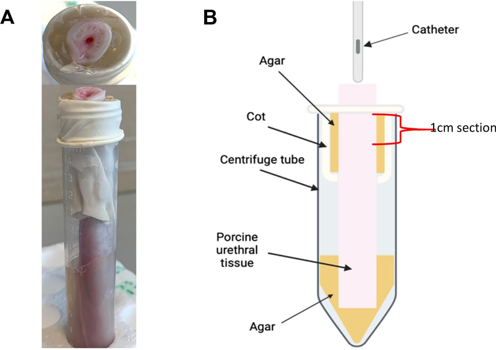
Burns J, Irwin RN, Quinn J et al (2025) An ex vivo porcine urethral model for investigating intermittent catheter-associated urethral microtrauma. Materials Design. 259:114727. https://doi.org/10.1016/j.matdes.2025.114727

Kato Y, Nagao Y (2012) Effect of polyvinylpyrrolidone on sperm function and early embryonic development following intracytoplasmic sperm injection in human assisted reproduction. Reprod Med Biol. 11:165-176. https://doi.org/10.1007/s12522-012-0126-9

Kazi S, Yang B, Carugo D, Stride E, Lavigne A (2025) A Novel Male Porcine Urethral Ex Vivo Model: Characterisation of Structural and Metabolic Viability. Abstract 657. ICS-EUS, Abu Dhabi. https://www.ics.org/2025/abstract/657 (accessed 31 October 2025)

Pollard D, Allen D, Irwin NJ, Moore JV, McClelland N, McCoy CP (2022) Evaluation of an Integrated Amphiphilic Surfactant as an Alternative to Traditional Polyvinylpyrrolidone Coatings for Hydrophilic Intermittent Urinary Catheters. Biotribology. 32:100223. https://doi.org/10.1016/j.biotri.2022.100223

TissueSource (2020) Advancing Urology Research with the Porcine Urinary Tissue Model. https://tissue-source.com/blog/urology-research/ (accessed 31 October 2025)