Bladder and ureters: an anatomy refresher

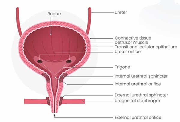
The bladder has two different roles which are both important to understand in relation to continence care. It is a hollow organ with stretchy walls which stores urine produced by the kidneys (Figure 1). Urine reaches the bladder via the ureters which connect it to the kidneys. The lining of the bladder is folded which allows it to expand to hold around 400-600ml of urine in healthy adults.

The bladder wall also contains detrusor muscles which allow it to perform its second role – the expulsion of urine. The muscles of the bladder contract during urination, alongside relaxation of the sphincters, to force urine out of the bladder via the urethra. These muscles play an important role in ensuring that the bladder empties fully. The fibres of the detrusor muscle often become enlarged in conditions that obstruct the urine outflow, such as benign prostatic hyperplasia, to compensate for the increased workload needed to empty the bladder (Jones, 2026).

The shape of the bladder changes depending on how full it is – it is an oval shape when full and flatter when empty. 

There are two sphincters in the urethra – the internal and external urethral sphincters. The internal urethral sphincter is thought to be purely functional in women (that is, it doesn’t have any muscle in it) and is thought to prevent semen from entering the bladder during ejaculation in men. In both sexes, the external urethral sphincter is made of skeletal muscle and is under voluntary control via the pudendal nerve.


Figure 1. Anatomy of the bladder.

Nerves supplying the bladder


The nervous control of the bladder is complicated, involving the sympathetic, parasympathetic and somatic parts of the nervous system.
  • The sympathetic system acts via the hypogastric nerve, and causes the detrusor muscle to relax, which helps the bladder to retain urine.
  • The parasympathetic system acts via the pelvic nerve. Signalling via this nerve causes the detrusor muscle to contract which stimulates urination.
  • The somatic system involves the pudendal nerve, which activates the external urethral sphincter and gives the person voluntary control over urination.

There are also sensory nerves in the bladder wall which send signals to the brain when the bladder becomes full, signalling the need to urinate.


Spinal cord injuries and the bladder


There are two different clinical syndromes that affect bladder function in people with spinal cord lesions, depending on whether the damage is below or above the base of the ribs (T12).


Reflex bladder – damage above T12


In this case, the nerve signals from the bladder wall are unable to reach the brain, and the patient will have no awareness of the bladder filling. There is also no control over the external urethral sphincter, so it is constantly relaxed. There is a functioning spinal reflex, where the parasympathetic system initiates detrusor contraction in response to bladder wall stretch. Thus, the bladder automatically empties as it fills – known as the reflex bladder.


Flaccid bladder – damage below T12


A spinal cord transection at this level will have damaged the parasympathetic outflow to the bladder. The detrusor muscle will be paralysed, unable to contract, and the spinal reflex does not function. In this case, the bladder fills uncontrollably, becoming abnormally distended until overflow incontinence occurs.

 

Links to incontinence


Different types of incontinence are caused by different problems with parts of the bladder.

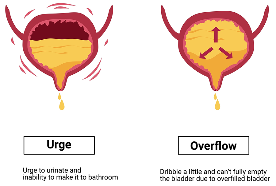
Urge incontinence is the sudden urge to urinate, which often results in bladder leakage. This occurs as a result of overactive contraction of the detrusor muscles.

Overflow incontinence occurs in people with dysfunction of the bladder outlet that stops the bladder from emptying completely. This is often seen in patients with prostatic hyperplasia, impaired detrusor contractility or narrowing of the urethra such as in cases of stricture disease, all of which can prevent the bladder from completely emptying. This causes the bladder to overfill with urine, resulting in a continuous leakage or dribbling of urine (Shermadou et al, 2023).


References


Jones O (2026) The urinary bladder https://teachmeanatomy.info/pelvis/viscera/bladder/ (accessed 6 May 2026)

Shermadou ES, Rahman S, Leslie SW (2023) Anatomy, abdomen and pelvis: bladder.  https://www.ncbi.nlm.nih.gov/books/NBK531465/ (accessed 5 May 2026)