How the anatomy of the spinal cord affects bladder function

The spinal cord controls bladder function through a network of sacral, lumbar, and thoracic nerve centres, along with the brainstem, that coordinate the storage and release of urine via signals from the brain (Shefchyk, 2002). Damage or variation along these spinal levels disrupts the reflex and voluntary balance, leading to specific patterns of bladder control issues.

Brainstem


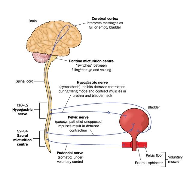
The pontine micturation centre in the brainstem (Figure 1) is a group of nerve cells found in the upper part of the pons. This acts as the main switch in the brain, controlling urination (micturation) by telling the bladder to empty and the urinary sphincters to relax (Rahman and Siddik, 2023). Other areas of the brainstem have been implicated in the coordination of urinary functions, including the locus coeruleus, the hypothalamus, the septal nuclei, and several cortical regions.
 

Thoracolumbar nerves


The hypogastric and pelvic nerves connect to the T10–L2 section of the spinal cord. Sympathetic pathways in these nerves suppress bladder contraction and close the internal sphincter in the bladder neck during urine storage.
 

Sacral nerves


The sacral part of this pathway involves the S2–4 section of the spinal cord. This acts as the core reflex centre (the sacral micturition centre); parasympathetic fibres prompt the bladder wall to squeeze, while the pudendal nerves control the external sphincter of the bladder. Urination results from the coordinated activity of sacral parasympathetic neurons and temporary inactivity of the α-motor neurons of the voluntary motor system (Purves et al, 2001).
 
Figure 1. Nervous control of the urinary bladder via the hypogastric, pelvic and pudendal nerves.
Figure 1. Nervous control of the urinary bladder via the hypogastric, pelvic and pudendal nerves. Credit: Blamb/Shutterstock


People with spinal cord injuries
 

People who are paraplegic, or people who have otherwise lost descending control of the sacral spinal cord, still have autonomic regulation of bladder function, since urination is eventually stimulated reflexively at the level of the sacral cord by distension of the bladder. Unfortunately, this reflex is not efficient if there is no descending motor control, resulting in a variety of problems in people who are paraplegic and others with diminished or absent central control of bladder function. The major issue faced is incomplete bladder emptying, which often leads to chronic urinary tract infections from the culture medium provided by retained urine (Purves et al, 2001). These people usually need to use a catheter to ensure that their bladder is drained sufficiently. 


Practice point


A recent study investigating the potential effects of polyvinylpyrrolidone (PVP) coating residues from hydrophilic ICs on male fertility found that exposure to these residues reduced sperm movement and caused abnormalities in sperm shape in a porcine model, although minimal damage to genetic material was observed. These findings raise potential concerns for men with spinal cord injuries who rely on IC (Irwin et al, 2026). Alternative catheter technologies, such as those incorporating an integrated amphiphilic surfactant (IAS), may be of benefit in this context. See here.
 

References


Irwin RN, Burns J, Quinn J, et al (2026) Investigating the effect of polyvinylpyrrolidone-based urinary catheter coatings on spermatozoa quality. Pharmaceutics and Pharmacology 5(1): rqag003. Available online: https://academic.oup.com/rpsppr/article/5/1/rqag003/8673208?login=false

Purves D, Augustine GJ, Fitzpatrick D et al, eds (2001) Autonomic Regulation of the Bladder. In: Neuroscience. 2nd edn. Sunderland (MA): Sinauer Associates. https://www.ncbi.nlm.nih.gov/books/NBK10886/ (accessed 21 August 2026)

Rahman M, Siddik AB (2023) Neuroanatomy, Pontine Micturition Center. Available online: www.ncbi.nlm.nih.gov/books/NBK557419/ (accessed 21 August 2026)

Shefchyk SJ (2002) Spinal cord neural organization controlling the urinary bladder and striated sphincter. Prog Brain Res 137: 71–82