Pelvic floor muscles: an anatomy refresher

 
The pelvic floor is important in terms of continence, so here we give a quick overview of what it is and why its function is so critical to both urinary and faecal continence. 

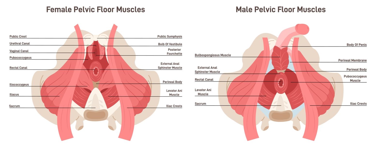
The pelvic cavity is the space inside the bones that make up the pelvis (Figure 1). A number of different muscles make up the wall of the pelvic cavity, with the pelvic floor muscles making up the bottom of the cavity. 

The pelvic floor is funnel shaped. It attaches to the walls of the pelvis and separates the contents of the pelvic cavity from the perineum.

There are gaps between the muscles in the pelvic floor which allow urination and defaecation - the urogenital hiatus is at the front of the pelvic floor and allows passage of the urethra (and the vagina in women), and the rectal hiatus, which is central and allows the rectum to pass through the muscles. 
 

What do the pelvic floor muscles do? 

The pelvic floor muscles have a number of important roles. They are used to support the organs that are in the abdomen and pelvis, and to resist increased pressure in the abdomen or pelvis, for example while you cough or if you are lifting heavy objects. They also act like a sphincter on the rectum and urethra, so relax to allow urination or defaecation (Fidoe, 2025).
Anatomy of female pelvis showing skeleton and muscles.
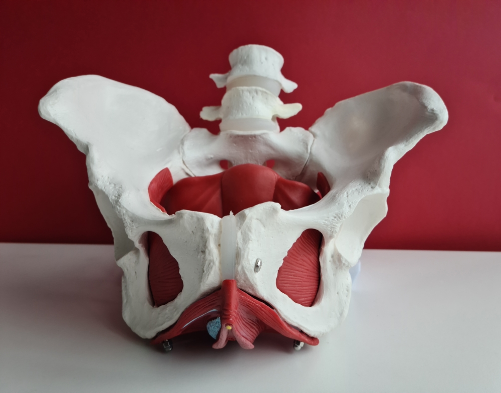
Figure 1. Anatomy of female pelvis showing skeleton and muscles.

Anatomy

There are three main components of the pelvic floor muscles (Figure 2): the levator ani muscles (largest), the coccygeus muscle and the fascia coverings of the muscles (Fidoe, 2025). 
 

Levator ani

The levator ani is a sheet of muscle which is made up of three separate pairs of muscles: 
  • Pubococcygeus
  • Puborectalis 
  • Iliococcygeus.
The puborectalis runs from the back of the pubis to attach on the opposite side. It forms a U-shaped sling around the anal canal and is the most important part of the levator ani in terms of maintaining faecal continence. Some fibres of the puborectalis form another U-shaped sling around the urethra or the urethra and vagina, and these fibres help to maintain urinary continence, particularly when the intra-abdominal pressure increases suddenly, ie when you sneeze. 

The puboccoygeus forms the largest part of the levator ani, and sits between the puborectalis and the iliococcygeus. Its role is to provide support for the pelvic and abdominal organs.   

The iliococcygeus is a thin muscle which sits at the back of the levator ani. It lifts the pelvic floor and the anorectal canal. 
 

Coccygeus

The coccygeus is a small triangular muscle which sits behind the levator ani muscles.  It supports the pelvic organs and can also be used to flex the coccyx (tail bone) (Fidoe, 2025). 
Anatomy of female and male pelvic floor muscles
Figure 2. Pelvic floor muscles. 
 

Pelvic floor dysfunction 

Pelvic floor dysfunction refers to a range of signs and symptoms that are related to abnormal functioning of the pelvic floor muscles. Weakening of these muscles can result in a loss of structural support to pelvic organs which can present as:
  • Urinary incontinence 
  • Faecal incontinence 
  • Genitourinary prolapse 
  • Pelvic pain 
  • Sexual dysfunction 
There are many causes of pelvic floor dysfunction, including trauma during childbirth, increasing age, obesity, and chronic straining.

Reference

Fidoe S (2025) The pelvic floor. https://teachmeanatomy.info/pelvis/muscles/pelvic-floor/ (accessed 7 May 2026)